Home Blog Page 4

Pseudomonas aeruginosa’s Five DppA Proteins Split Peptide Duties in Unexpected Ways

0
Pseudomonas aeruginosa’s Five DppA Proteins Split Peptide Duties in Unexpected Ways source image 1
Source article image 2
Source article image 3
Source article image 4
Source article image 5

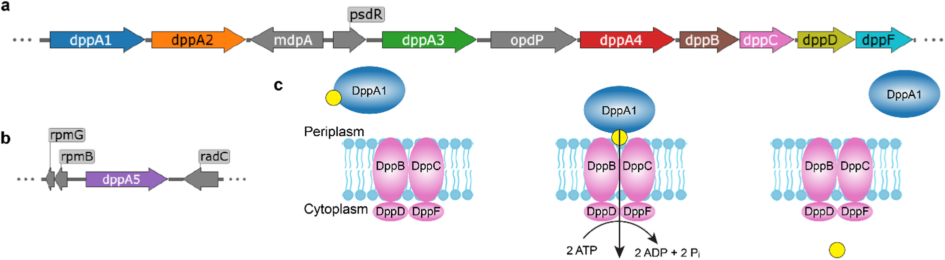
Pseudomonas aeruginosa is a notorious opportunistic pathogen, and part of its survival strategy is a remarkably flexible nutrient uptake network. One of the key systems it uses is the dipeptide permease, or Dpp, which imports short peptides from the periplasm into the cell. What makes this system unusual is that, unlike the single solute-binding protein seen in many bacteria, P. aeruginosa encodes five DppA paralogs.

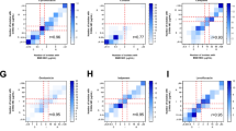
A new study has now mapped the binding preferences of all five proteins, revealing that they are far from interchangeable. Using differential scanning fluorimetry and a library of 281 di- and tripeptides, the researchers found clear specialization among the paralogs. DppA1 and DppA3 showed a stronger preference for dipeptides, while DppA2 and DppA4 were more responsive to tripeptides. DppA5, by contrast, showed no detectable peptide binding under the conditions tested.

The results suggest that the multiple DppA proteins do not simply duplicate one another’s function. Instead, they appear to broaden the range of peptide substrates that P. aeruginosa can capture from its environment. That flexibility may help the bacterium thrive in nutrient-poor or competitive settings, including the human host.

The work also reinforces the idea that peptide transport in this pathogen may be tied to more than metabolism alone. Dpp-related proteins have previously been linked to behaviors such as biofilm formation and chemotaxis, raising the possibility that ligand recognition by DppA proteins could influence signaling as well as uptake.

From a practical standpoint, the study offers two important takeaways. First, it corrects the assumption that the five paralogs are functionally redundant. Second, it provides a more detailed foundation for thinking about Dpp as a potential route for peptide-based drug delivery strategies, including Trojan horse antimicrobials designed to exploit bacterial transport systems.

In short, P. aeruginosa appears to use a diversified peptide-sensing toolkit to expand what it can import, and that diversification may be part of what makes this pathogen such a resilient problem in clinical settings.

A Faster, Additive-Free Native Chemical Ligation Strategy Using Vinyl Thianthrenium Tetrafluoroborate

0
A Faster, Additive-Free Native Chemical Ligation Strategy Using Vinyl Thianthrenium Tetrafluoroborate source image 1
Source article image 2
Source article image 3

New route for faster peptide ligation

Native chemical ligation (NCL) remains one of the most important reactions in chemical protein synthesis because it joins peptide fragments with high selectivity and preserves the native peptide bond. But the classic approach often depends on peptide thioesters that can be slow to react, pushing chemists to use large amounts of thiol additives to keep the reaction moving.

A new study introduces a different strategy: using vinyl thianthrenium tetrafluoroborate (VTT) to rapidly activate fully unprotected peptide thioacids. In the presence of VTT, these thioacids are converted quantitatively into reactive thioester intermediates, which then undergo native chemical ligation with N-terminal cysteinyl peptides. The process is reported to be fast, chemoselective, and free of the usual additive burden.

Why this matters for protein synthesis

One of the main advantages of the new method is compatibility with one-pot workflows. Traditional NCL procedures often require a thiol additive such as MPAA, but those additives can interfere with downstream radical desulfurization steps used to convert cysteine into alanine. That creates extra purification or handling steps.

By avoiding additive use, the VTT-enabled ligation is better suited to streamlined NCL-desulfurization sequences. According to the authors, the method works without epimerization and supports efficient post-ligation transformations in the same vessel.

Demonstrated on complex targets

The researchers showed the utility of the platform by synthesizing hyalomin-3 from two fragments through a one-pot thioesterification-ligation-desulfurization sequence. They also used the approach in a more complex multisegment assembly of ubiquitin, completed through a one-pot C-to-N sequential three-segment condensation strategy.

These examples suggest that the chemistry could simplify access to proteins that are difficult to build using standard peptide assembly methods, especially where speed, cleaner reaction profiles, and operational simplicity are important.

Broader implications

For peptide and protein chemists, the appeal of this work is straightforward: faster activation, fewer additives, and greater compatibility with downstream editing steps. If the method proves broadly general across more sequences and protein contexts, it could become a useful addition to the chemical protein synthesis toolkit.

As CPS continues to expand into the preparation of modified proteins, probes, and therapeutic research tools, methods that reduce steps and improve one-pot efficiency are likely to draw strong interest.

A UV Crosslinking Strategy Reveals a Hidden Ligand for Orphan GPCR GPR50

0
A UV Crosslinking Strategy Reveals a Hidden Ligand for Orphan GPCR GPR50 source image 1
Source article image 2
Source article image 3

Orphan G protein-coupled receptors remain one of the most challenging classes of signaling proteins to study because their natural ligands are unknown. A new method described in Nature Chemical Biology offers a way to tackle that problem by using site-specific UV-mediated crosslinking to trap receptor-ligand interactions long enough to identify them.

Using this approach, researchers identified the neuropeptide Little-LEN as an agonist for GPR50, an orphan GPCR. The finding adds a missing piece to the biology of a receptor previously lacking a defined endogenous partner and suggests a role in tuning energy expenditure according to metabolic state.

Why orphan GPCRs matter

GPCRs are central to cell communication and are major drug targets, but orphan receptors have no assigned natural ligand, making them difficult to place in physiology. Finding those ligands can uncover new signaling pathways and open the door to therapeutic targeting.

How UV crosslinking helps

The key advance is the use of a light-triggered, site-specific crosslinking strategy. In practice, this means a ligand can be positioned so that, when exposed to UV light, it forms a covalent link with its receptor at the contact site. That stable capture makes it easier to pull down and identify the interaction.

Compared with conventional screening approaches, this kind of chemical trapping can be especially useful for transient or low-affinity interactions that might otherwise escape detection.

A peptide clue to metabolic regulation

The identification of Little-LEN as a GPR50 agonist is notable not only because it de-orphans a receptor, but also because it connects the receptor to peptide signaling in metabolism. The reported relationship between GPR50 and energy expenditure suggests the receptor may help organisms adapt their energy use to nutritional state.

For peptide researchers, the result is a reminder that small endogenous peptides can still hide in plain sight, waiting for better tools to expose their targets. For GPCR biology, it is another example of how modern chemical methods can turn an orphan receptor into a defined signaling pathway.

What comes next

Methods like this could accelerate ligand discovery across the orphan GPCR family, especially where traditional deconvolution strategies have stalled. As more receptors are matched with their native ligands, the field should gain a clearer view of how peptide signals shape physiology, metabolism, and disease.

Deep Learning Helps Design Better Protease Substrates

0
Deep Learning Helps Design Better Protease Substrates source image 1
Source article image 2
Source article image 3
Source article image 4
Source article image 5

Designing peptide substrates for proteases has always been a difficult mix of biology, chemistry, and brute-force screening. The challenge is not just finding a sequence a protease can cut, but finding one that is cut efficiently and selectively. With an enormous sequence space and limited experimental throughput, substrate discovery has often relied on literature mining, expert intuition, or expensive large-scale screens.

A new study introduces CleaveNet, an end-to-end deep learning pipeline built to address that bottleneck. Rather than only predicting whether a peptide will be cleaved, the system is designed to generate candidate substrates directly. In other words, it aims to move from passive prediction to active design.

The method was applied to matrix metalloproteinases, a family of proteases with important roles in tissue remodeling, inflammation, and cancer. According to the study, CleaveNet can produce peptide substrates with favorable biophysical properties while also capturing known cleavage preferences and uncovering motifs that had not been well characterized before.

One notable feature is a conditioning tag that allows users to steer generation toward desired cleavage behavior. That means the model can be guided to favor more efficient substrates or, importantly, more selective ones. Selectivity matters because closely related proteases often recognize overlapping peptide patterns, making it hard to design probes that respond to just one target.

The researchers validated the generated peptides in a large in vitro screen, including the especially demanding case of designing selective substrates for MMP13. Experimental results supported the idea that AI-assisted design can reduce the trial-and-error burden normally associated with protease substrate development.

For peptide researchers, the broader significance is clear: if models like CleaveNet continue to improve, they could shorten the path from enzyme biology to usable probes, diagnostic triggers, and therapeutic tools. The approach may also point toward a more general future for enzyme engineering, where deep learning helps explore chemical space that is simply too large to search by hand.

In a field where substrate design has long been constrained by scale and specificity, this work suggests that generative AI may become a practical discovery engine rather than just an analytical aid.

A UV Crosslinking Strategy Reveals a Ligand for Orphan GPCR GPR50

0
A UV Crosslinking Strategy Reveals a Ligand for Orphan GPCR GPR50 source image 1
Source article image 2
Source article image 3

Orphan G protein-coupled receptors remain some of the most elusive drug targets in biology because their natural ligands are often unknown. A new approach reported in Nature Chemical Biology uses site-specific UV-mediated crosslinking to help solve that problem, providing a way to capture receptor–peptide interactions that can be difficult to detect with conventional screening.

Using this strategy, the researchers identified Little-LEN, a neuropeptide, as an agonist for GPR50. That finding is notable not only because it assigns a ligand to a previously orphan receptor, but also because it connects the pair to regulation of energy expenditure in response to metabolic state.

The basic idea is elegant: introduce a light-activated crosslinking handle at a defined position so that, when the right ligand binds, the interaction can be trapped and analyzed. This kind of method could be especially useful for peptide ligands, which often act through transient, selective contacts and may be missed by broader discovery workflows.

For peptide science, the result highlights two important themes. First, many endogenous signaling peptides likely still have undiscovered receptor partners. Second, chemical biology tools that preserve native-like binding while adding a controllable capture step can accelerate receptor deorphanization. In the long run, that could improve our understanding of neuroendocrine signaling and point to new therapeutic opportunities in metabolic regulation.

GPR50 now joins the growing list of orphan receptors with clearer biological context, and Little-LEN adds another example of how peptide ligands can illuminate previously hidden signaling pathways.

New DNA-Peptide Conjugation Strategy Boosts Nuclear Delivery in Non-Dividing Cells

0
New DNA-Peptide Conjugation Strategy Boosts Nuclear Delivery in Non-Dividing Cells source image 1
Source article image 2
Source article image 3
Source article image 4
Source article image 5

Delivering DNA into the nucleus remains one of the biggest bottlenecks in non-viral gene therapy. Unlike mRNA, DNA has to cross the nuclear barrier before it can be transcribed, and that step is especially difficult in cells that are not actively dividing. A new study describes a peptide-based strategy designed to make that journey more efficient.

The researchers built a modular workflow for creating DNA-peptide conjugates using an enzyme-driven tagging approach. Their platform produces peptide-modified DNA gene cassettes, referred to as DNA-PepTAG, by attaching nuclear localization signal (NLS) peptides to linear DNA. The goal is to improve how effectively DNA cargo reaches the nucleus after delivery.

In growth-arrested cells, gene cassettes carrying NLS peptides showed stronger nuclear localization, higher mRNA output, and greater reporter expression than unmodified DNA. In tests using an eGFP reporter delivered by lipofection, expression increased by up to about 10-fold in some conditions.

The team also screened several different NLS sequences across multiple human cell lines and found that the best-performing peptide could depend on the cell type. Two NLS peptides, PLSCR-1 and extSV40, stood out for producing consistently strong results across the models tested, suggesting they may be useful starting points for broader applications.

Importantly, the approach was not limited to a single payload size or protein class. The system was used to deliver gene cassettes encoding both cytosolic and secreted proteins, and it was tested across DNA constructs ranging from 1.3 kbp to 7 kbp. That range suggests the method may be adaptable to a variety of therapeutic or research applications.

Why does this matter? Many current DNA delivery methods rely on high doses to overcome poor nuclear entry, which can increase toxicity and complicate translation. If peptide-guided nuclear delivery can improve expression with less DNA, it could make non-viral gene delivery more practical for future therapies.

As with many delivery technologies, the details matter. The study reinforces that the identity of the peptide, the way it is attached, and the cell context all influence performance. Still, the new conjugation workflow offers a more controlled route to DNA-NLS engineering and may help address a long-standing obstacle in DNA therapeutics.

Overall, the work points to peptide-tagged DNA as a promising platform for boosting nuclear delivery in non-dividing cells, with potential relevance for gene therapy and other DNA-based applications.

A modular peptide-DNA conjugation strategy boosts nuclear delivery in non-dividing cells

0
A modular peptide-DNA conjugation strategy boosts nuclear delivery in non-dividing cells source image 1
Source article image 2
Source article image 3
Source article image 4

Getting DNA into the nucleus is one of the biggest bottlenecks in non-viral gene delivery. That challenge becomes even more pronounced in growth-arrested cells, where the usual route of nuclear entry during cell division is largely unavailable. In a new study, researchers describe a modular peptide-conjugation workflow that helps DNA cargo cross this barrier more effectively.

The team built peptide-modified gene cassettes by first generating DNA oligonucleotide-peptide conjugates with an enzymatic tagging system based on E. coli tRNA guanine transglycosylase, then ligating those units onto linear DNA. The result was a DNA-peptide platform, referred to as DNA-PepTAG, designed to present nuclear localization signal (NLS) peptides in a more controlled and reproducible way.

When the researchers delivered an eGFP reporter by lipofection into growth-arrested cells, NLS-modified cassettes showed stronger nuclear localization, higher mRNA output, and greater protein expression than unmodified DNA. In the best cases, expression increased by roughly an order of magnitude.

One important finding was that not all NLS peptides performed equally across cell types. Screening in several human growth-arrested cell lines revealed clear cell-type preferences for DNA nuclear transport. Still, two candidates stood out: PLSCR-1 and extSV40 produced consistently strong expression across the tested systems, suggesting they may be particularly useful for broader applications.

The approach was also tested with different payloads, including genes encoding both cytosolic and secreted proteins, as well as gene cassettes ranging from about 1.3 kbp to 7 kbp. This range suggests the method is not limited to a single reporter construct and may be adaptable to a variety of therapeutic or research DNA cargos.

For peptide researchers, the study is a useful reminder that peptide identity is only part of the story. How a peptide is attached to DNA can strongly influence whether it can do its job. By offering a site-specific, modular conjugation strategy, this platform may help make DNA-NLS delivery more reliable and more dose-efficient.

The broader implication is clear: if DNA therapeutics are to compete in non-viral delivery, the field will likely need better tools for guiding cargo into the nucleus. DNA-PepTAG adds a promising option to that toolkit.

A new way to shut down the ATAC histone acetyltransferase complex

0
A new way to shut down the ATAC histone acetyltransferase complex source image 1
Source article image 2
Source article image 3
Source article image 4

Histone acetyltransferases help turn genes on by adding acetyl groups to chromatin, but these enzymes do not work in isolation. They operate as part of multi-protein assemblies that can change where they bind, which substrates they prefer, and what biological programs they control. That complexity has made it difficult to study one complex without disturbing others that use the same core catalytic machinery.

A new study takes a different route. Instead of inhibiting the shared HAT enzyme, the researchers designed chemical inhibitors against YEATS2, a component that is specific to the ATAC coactivator complex. This approach allowed them to selectively weaken ATAC function while leaving other HAT-containing complexes largely untouched.

The lead compound, LS-170, reduced how much ATAC was retained on chromatin, lowered ATAC-linked histone acetylation, and changed the expression of genes regulated by the complex. In cells and in a lung cancer mouse model, those effects translated into a measurable reduction in tumor growth.

The work is notable for two reasons. First, it provides a chemical tool for dissecting the roles of a single HAT complex in gene regulation. Second, it suggests that complex-selective targeting may be a useful therapeutic strategy, especially in cancers that depend on ATAC-driven transcriptional programs.

For peptide and chromatin-focused drug discovery, the study reinforces an important idea: sometimes the best way to modulate an enzyme pathway is not to hit the active site, but to target a partner protein that gives the complex its unique biology.

Structural and sequencing data from the project have also been deposited publicly, supporting follow-up work on the ATAC complex and its inhibitors.

A Complex-Specific Strategy to Block the ATAC Histone Acetyltransferase Complex

0
A Complex-Specific Strategy to Block the ATAC Histone Acetyltransferase Complex source image 1
Source article image 2

Histone acetyltransferases help control gene expression by loosening chromatin and making DNA more accessible to the transcriptional machinery. But these enzymes rarely work in isolation. Instead, they act within multi-protein complexes that shape where they go in the genome, which substrates they modify, and how they influence cell behavior.

That complexity creates a challenge for drug discovery. If a compound blocks the shared catalytic enzyme, it may hit several related complexes at once, making it difficult to tease apart which biological effects come from which assembly. In a new study, researchers took a more selective approach: rather than targeting the common HAT enzyme, they designed inhibitors against YEATS2, a subunit unique to the ATAC complex.

The goal was to disrupt ATAC specifically and observe what happens when this complex is removed from chromatin. The lead compound, LS-170, showed the strongest activity. According to the study, it reduced ATAC occupancy on chromatin, lowered ATAC-dependent histone acetylation, and suppressed expression of genes controlled by ATAC.

Those molecular changes also translated into a functional effect in vivo. In a lung cancer mouse model, treatment with the inhibitor significantly restrained tumor growth, suggesting that ATAC may be a promising therapeutic vulnerability in at least some cancer settings.

Beyond the therapeutic angle, the work provides a useful chemical tool for studying how HAT complexes behave in cells. Because ATAC is part of a broader family of acetyltransferase assemblies with overlapping enzymatic features, a complex-selective inhibitor could help researchers map out which genes and pathways depend on ATAC specifically, rather than on HAT activity in general.

The study also adds to a growing view in chromatin biology: the composition of a protein complex can be just as important as the enzyme it carries. By targeting a complex-specific subunit, the authors demonstrate a way to dissect chromatin regulation with greater precision and potentially open a new route for epigenetic drug development.

Structural data associated with the work were deposited in the Protein Data Bank, and sequencing datasets were made publicly available through GEO.

Nature publishes correction to cathepsin D peptide probe article

0
Nature publishes correction to cathepsin D peptide probe article

Nature Communications Chemistry has issued an author correction for the article on a fluorogenic peptide-based probe designed to detect Cathepsin D in macrophages.

The correction addresses a set of figure and supplementary note artwork errors. In the original version, the CatD-P3 probe was drawn with an incomplete PEG tag, the click-chemistry reagent was shown with the wrong alkyne depiction, and the PEG reagent used in the synthesis was also illustrated incorrectly in the figures and supplementary material. A separate labeling issue in one figure misplaced charges within the BODIPY fluorophore.

The publisher states that these problems were visual only and do not alter the experimental results or the paper’s conclusions. The HTML and PDF versions have been updated to include the corrected versions of Figures 1 and 2 and Supplementary Note 4.

The corrected article remains open access under a Creative Commons Attribution 4.0 International License.

AI generated illustration

AI-generated illustration